
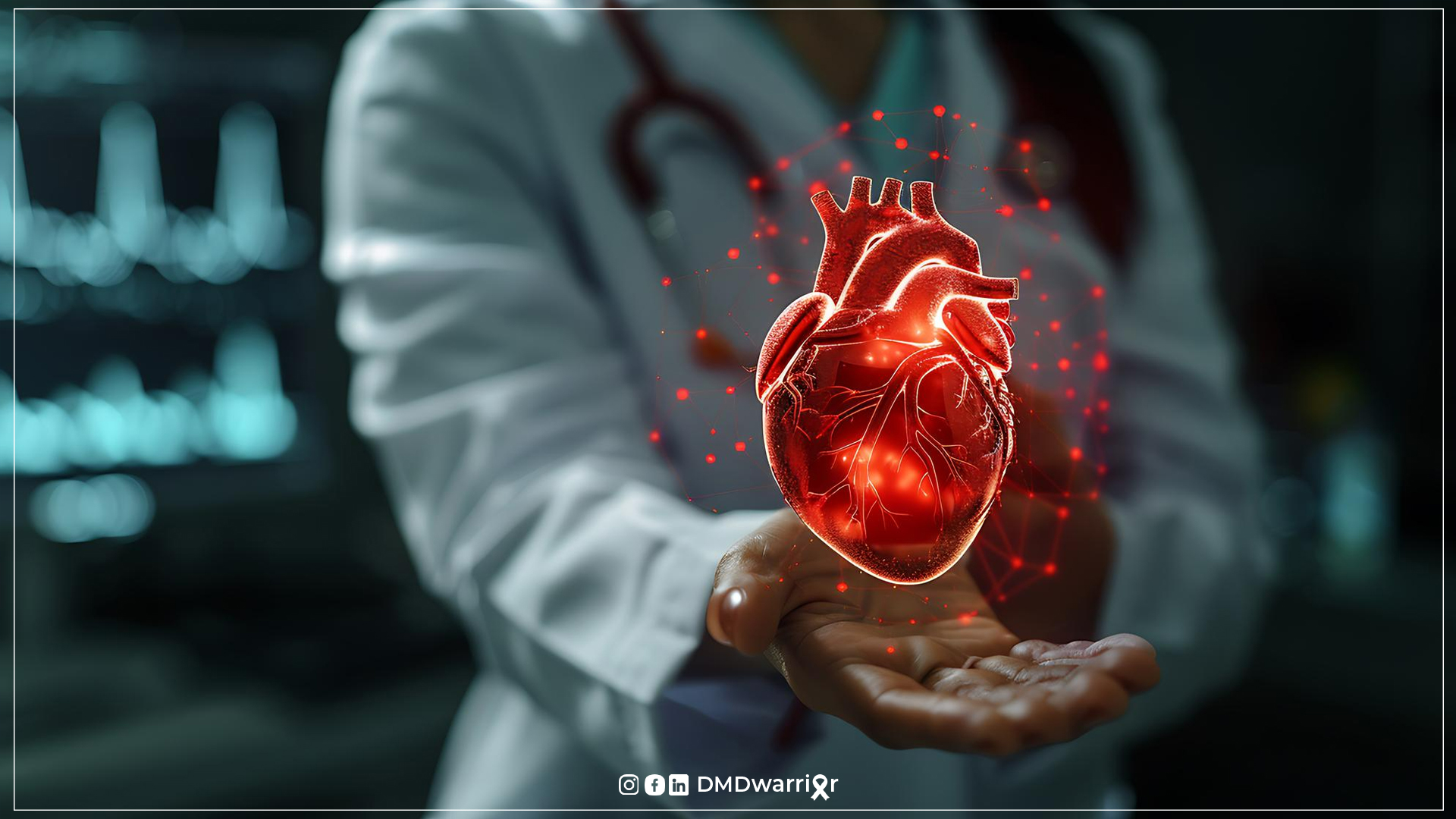
Heart health in Duchenne Muscular Dystrophy is a critical yet often underrecognized aspect of managing this progressive condition. As advances in care continue to extend life expectancy, cardiomyopathy has emerged as a leading concern, making early detection and proactive treatment strategies more important than ever.
Understanding how Duchenne Muscular Dystrophy affects the heart—and what modern medicine can do to slow its progression—empowers patients, caregivers, and clinicians to take timely action.
This article provides an in-depth, guide to heart health in Duchenne muscular dystrophy, including causes, early detection, modern imaging techniques, and evidence-based treatment strategies.

Table of Contents
Understanding Heart Health in Duchenne Muscular Dystrophy
The hallmark of Duchenne muscular dystrophy is the absence of dystrophin, a protein that helps maintain the stability of muscle cells. Without dystrophin, muscle fibers become fragile and are progressively replaced by fat and fibrotic tissue.
Although skeletal muscles are most visibly affected, the heart is also a muscle. Over time, dystrophin deficiency causes damage to cardiac muscle cells, leading to structural changes and impaired heart function.
This process results in Duchenne muscular dystrophy cardiomyopathy, a condition characterized by progressive weakening of the heart muscle.
Research shows that most individuals with DMD develop some form of cardiac involvement by adolescence or early adulthood. Cardiac complications can include:
- Dilated cardiomyopathy
- Cardiac Arrhythmia
- Reduced cardiac output
- Progressive heart failure
A large systematic review of over 9,000 patients found that dystrophin deficiency leads to myocardial fibrosis, left ventricular dysfunction, and arrhythmias, ultimately contributing to heart failure in many patients with DMD.1
Cardiac Involvement in Duchenne Muscular Dystrophy
Why the Heart Is Affected
Cardiac involvement in Duchenne muscular dystrophy develops because dystrophin plays an essential role in maintaining the structural integrity of heart muscle cells.
Without dystrophin:
- The cell membrane becomes fragile
- Calcium leaks into the cells
- Cells undergo degeneration and death
- Fibrotic tissue replaces normal muscle
This progressive damage leads to weakening and enlargement of the heart chambers, particularly the left ventricle.
Symptoms of Heart Problems in Duchenne Muscular Dystrophy
One of the challenges in managing heart problems in Duchenne muscular dystrophy is that symptoms may be subtle or absent in early stages.
Common symptoms include:
- Fatigue
- Shortness of breath
- Rapid heartbeat
- Swelling of the legs or abdomen
- Reduced exercise tolerance
However, symptoms can be difficult to recognize because patients with DMD already have limited mobility.
Studies indicate that cardiac dysfunction symptoms are often underrecognized due to severe physical impairment associated with the disease.
Dilated Cardiomyopathy in DMD
The most common cardiac complication in DMD is dilated cardiomyopathy in DMD.
Dilated cardiomyopathy occurs when:
- The heart chambers enlarge
- The heart muscle becomes thin and weak
- The pumping ability of the heart declines
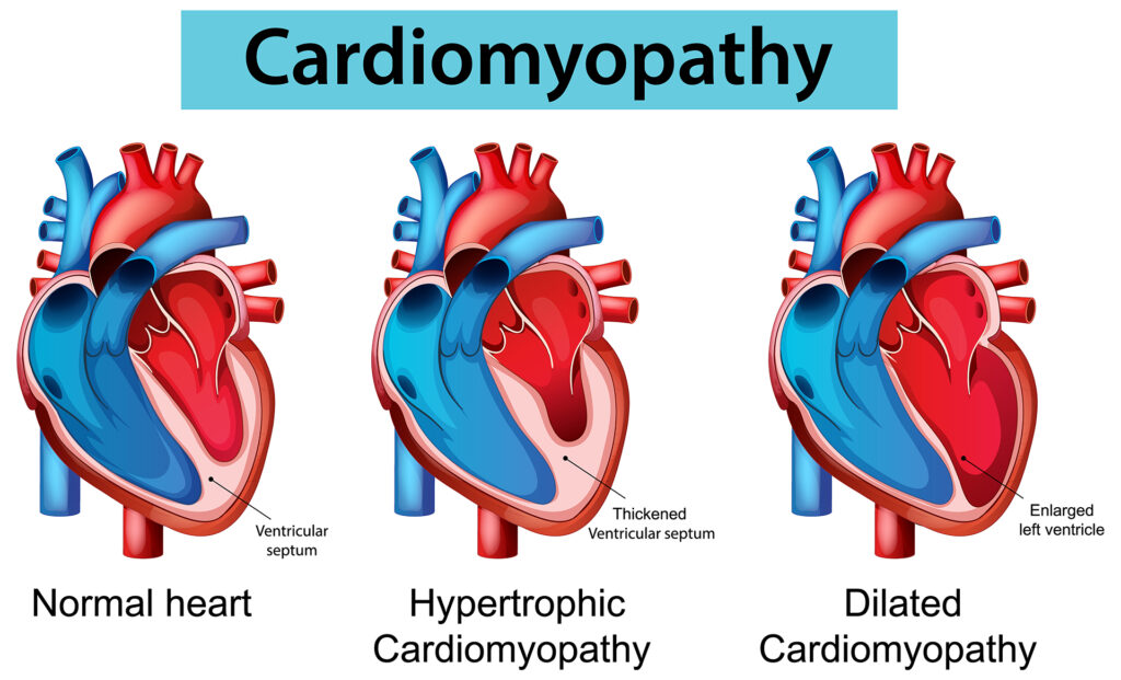
Over time, this leads to reduced left ventricular ejection fraction (LVEF)—a key measurement of heart function.
Timeline of Cardiomyopathy Development
Research suggests:
- Cardiomyopathy may begin as early as age 10
- Most patients with DMD develop cardiomyopathic features between ages 10 and 15 years.
- Nearly all adult patients develop some degree of cardiomyopathy
One study reported that the average age of onset of cardiomyopathy in DMD is approximately 14–15 years.
Because cardiomyopathy often begins before symptoms appear, early monitoring is essential.
Early Detection of Cardiomyopathy in DMD
Early detection of cardiomyopathy in DMD is critical because early treatment can slow disease progression.
Recommended Screening Guidelines
Current international care guidelines recommend:
- Baseline cardiac evaluation at diagnosis
- Annual cardiac monitoring
- More frequent monitoring if abnormalities are detected
Regular screening helps physicians identify early signs of cardiac dysfunction before symptoms develop.
Common screening tools include:
- Echocardiography
- Cardiac MRI
- Electrocardiography (ECG)
- Biomarker testing
Cardiac MRI in Duchenne Muscular Dystrophy
One of the most powerful tools for detecting early heart damage in DMD is cardiac MRI in Duchenne muscular dystrophy.
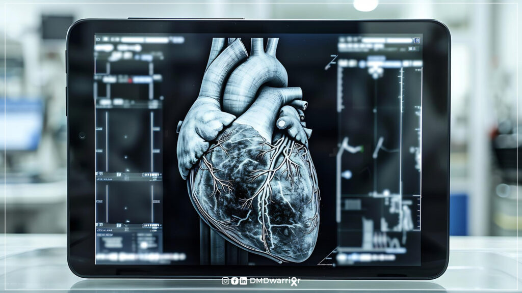
Cardiac MRI offers several advantages over traditional echocardiography:
Benefits of Cardiac MRI
- Detects early myocardial fibrosis
- Provides detailed imaging of heart muscle
- Identifies structural changes before symptoms appear
- Measures heart function accurately
Advanced MRI techniques such as late gadolinium enhancement (LGE) can detect scar tissue in the heart muscle, allowing physicians to identify early cardiomyopathy.
Studies show that MRI can detect heart damage years before clinical symptoms or echocardiography abnormalities appear.
Cardiac Management in Duchenne Muscular Dystrophy
Effective cardiac management in Duchenne muscular dystrophy requires a multidisciplinary approach involving:
- Cardiologists
- Neurologists
- Pulmonologists
- Genetic specialists
- Rehabilitation professionals
Management strategies typically include:
- Regular monitoring
- Preventive medications
- Lifestyle adjustments
- Advanced therapies
A coordinated care team is essential because cardiac and respiratory complications are the leading causes of mortality in DMD.2
Discover More: Multidisciplinary Neuromuscular Team in Duchenne
Treatment of Duchenne Muscular Dystrophy Cardiomyopathy
The treatment of Duchenne muscular dystrophy cardiomyopathy focuses on:
- Slowing disease progression
- Improving heart function
- Preventing heart failure
- Enhancing quality of life
Modern treatment strategies include:
- ACE inhibitors
- Beta-blockers
- Antimineralcorticoids
- Advanced heart failure therapies
Treatment with angiotensin converting enzyme (ACE) inhibitors, angiotensin receptor blockers (ARB), beta blocker, aldosterone antagonists, and SGLT-2 inhibitors can slow the course of cardiac muscle deterioration in DMD if the medications are started early. Some data suggest that corticosteroids may delay the onset of cardiomyopathy.3
Evidence suggests that early pharmacological therapy significantly improves cardiac outcomes in DMD.
ACE Inhibitors and Beta-Blockers for DMD Heart Disease
Among the most widely used treatments are ACE inhibitors and beta-blockers for DMD heart disease.
How ACE Inhibitors Work
ACE inhibitors:
- Lower blood pressure
- Reduce cardiac workload
- Prevent heart remodeling
- Slow progression of cardiomyopathy
Common examples include:4
- Enalapril
- Perindopril
- Lisinopril
A randomized clinical trial evaluating enalapril combined with a beta-blocker suggested that early therapy may delay the progression of left ventricular dysfunction in DMD patients.5
Benefits of Beta-Blockers
Beta-blockers:
- Slow heart rate
- Reduce stress on the heart
- Improve cardiac function
- Lower arrhythmia risk
Long-term studies show that continuous treatment with ACE inhibitors and beta-blockers is associated with a reduced decline in left ventricular ejection fraction.2
Preventing Heart Failure in Duchenne Muscular Dystrophy
If cardiomyopathy progresses without treatment, it may lead to heart failure in Duchenne muscular dystrophy.
Heart failure occurs when the heart can no longer pump blood effectively to meet the body’s needs.
Symptoms may include:
- Severe fatigue
- Fluid buildup
- Breathing difficulties
- Reduced oxygen delivery to tissues
Preventive strategies include:
- Early medication
- Routine cardiac monitoring
- Early treatment of arrhythmias
- Respiratory support
Research suggests that initiating therapy as soon as cardiac abnormalities appear significantly improves long-term outcomes.7
Emerging Therapies and Future Research
Advances in cardiology and neuromuscular medicine are opening new possibilities for treating Duchenne cardiomyopathy.
Emerging therapies include:
Mineralocorticoid receptor antagonists
Drugs like eplerenone may reduce myocardial fibrosis.
Gene therapy
Experimental therapies aim to restore dystrophin expression.
Cardiac regenerative therapies
Stem cell approaches are under investigation.
Advanced heart failure treatments
These may include:
- Mechanical circulatory support
- Implantable cardiac devices
- Heart transplantation in selected patients
New research also suggests that combination therapies targeting multiple cardiac pathways may improve outcomes.
Lifestyle and Supportive Strategies for Heart Health
Although medications are essential, lifestyle and supportive care can also help protect heart health.
Families and caregivers should focus on:
Regular follow-up with specialized neuromuscular centers is strongly recommended.
How often should you have your heart checked in Duchenne?
At Diagnosis
- A baseline cardiac evaluation should be performed as soon as Duchenne is diagnosed.
- This typically includes:
- Electrocardiogram (ECG)
- Echocardiogram
- Sometimes Cardiac MRI
Ages 0–9
- Heart check every 1–2 years
- Usually performed with an echocardiogram and ECG.
Age 10 and Older
- Heart check at least once every year
- This is when cardiac involvement in Duchenne muscular dystrophy commonly begins.
If Heart Changes Are Detected
- Monitoring may increase to every 6 months.
- Doctors may add medications such as ACE inhibitors or beta‑blockers to slow progression.
Cardiac MRI Monitoring
Many specialists recommend cardiac MRI every 1–2 years after age 10, because it can detect early heart muscle damage before it appears on ultrasound.
Before surgery
People with Duchenne muscular dystrophy should always have their heart checked before surgery. Because Duchenne often causes cardiac involvement and cardiomyopathy, a pre-operative cardiac evaluation helps doctors reduce anesthesia and surgical risks. Look Here: Surgery Under Anesthesia
FAQ: Heart Health in Duchenne Muscular Dystrophy
When do heart problems start in Duchenne muscular dystrophy?
Heart complications typically begin in adolescence, although subtle changes may appear earlier. Most patients develop cardiomyopathy during their teenage years.
Why does Duchenne muscular dystrophy affect the heart?
DMD is caused by a mutation in the dystrophin gene. Without dystrophin, cardiac muscle cells become fragile and gradually deteriorate. Read More: What is Dystrophin Gene?
What is cardiomyopathy in Duchenne muscular dystrophy (DMD)?
Cardiomyopathy in Duchenne muscular dystrophy refers to the progressive weakening of the heart muscle caused by the absence of dystrophin. Over time, this leads to dilated cardiomyopathy, reduced left ventricular function, and eventual heart failure. Cardiac involvement is almost universal in DMD and often develops silently before symptoms appear.
How is cardiomyopathy detected in DMD?
Doctors use several tests:
• Echocardiography
• Cardiac MRI
• Electrocardiogram (ECG)
• Blood biomarkers
Cardiac MRI is particularly useful for detecting early heart damage
What medications are used to treat DMD cardiomyopathy?
Common medications include:
• ACE inhibitors
• Beta-blockers
• Antimineralcorticoids
These drugs help slow disease progression.
Can heart problems in DMD be prevented?
While they cannot be completely prevented, early treatment and regular monitoring can significantly delay progression.
Is cardiac MRI necessary for DMD patients?
Yes. Many specialists recommend periodic cardiac MRI because it detects heart damage earlier than other imaging methods.
How can early cardiac involvement in DMD be detected?
Early cardiac involvement in DMD is typically asymptomatic, making proactive screening essential. Detection methods include:
• Echocardiography (ECHO) to assess heart function
• Cardiac MRI (CMR) to identify fibrosis (scar tissue) before functional decline
• Electrocardiogram (ECG) for rhythm abnormalities
Cardiac MRI is particularly sensitive in detecting early myocardial damage, even when ECHO results appear normal.
Why are ACE inhibitors used in DMD cardiomyopathy?
ACE inhibitors are a cornerstone of cardiac treatment in DMD because they:
• Reduce cardiac workload (afterload reduction)
• Slow progression of left ventricular dysfunction
• Delay onset of symptomatic heart failure
They are often initiated prophylactically, even before measurable cardiac dysfunction appears, based on clinical guidelines.
How often should ECHO and cardiac MRI be performed in DMD?
Cardiac monitoring frequency depends on age and disease stage:
ECHO:
• Typically every 6–12 months, starting at diagnosis or early childhood
Cardiac MRI:
• Usually every 1–2 years, or earlier if abnormalities are suspected
MRI is increasingly preferred for long-term monitoring due to its higher sensitivity for fibrosis detection.
How common are heart rhythm disorders in DMD?
Cardiac arrhythmias are relatively common in DMD, especially in later stages of the disease. These may include:
• Sinus tachycardia
• Ventricular arrhythmias
• Conduction abnormalities
The risk increases as cardiomyopathy progresses, making regular rhythm monitoring essential.
What should a cardiology follow-up plan look like in DMD?
A comprehensive cardiology follow-up plan for DMD should include:
• Routine imaging (ECHO and MRI)
• Regular ECG or Holter monitoring
• Early initiation of cardioprotective medications (ACE inhibitors, beta-blockers)
• Multidisciplinary coordination with neurology and pulmonology
Follow-up should ideally begin at diagnosis and continue lifelong.
What are the side effects of heart medications used in DMD?
Common medications and their potential side effects include:
• ACE inhibitors:
Low blood pressure, dizziness, kidney function changes, dry cough
• Beta-blockers:
Fatigue, slow heart rate, cold extremities
• Mineralocorticoid receptor antagonists (e.g., eplerenone):
Elevated potassium levels, kidney function changes
Regular monitoring helps manage these risks effectively
Is there a risk of sudden cardiac events in DMD?
Yes, there is a risk of sudden cardiac events in Duchenne muscular dystrophy, particularly in advanced stages with severe cardiomyopathy or arrhythmias. However, with:
• Early cardiac screening
• Timely medical therapy
• Close cardiology follow-up
this risk can be significantly reduced.
Learn More: Cardiovascular Disease in Duchenne Muscular Dystrophy
Conclusion
Heart health has become a central component of Duchenne muscular dystrophy care. As treatments extend life expectancy, managing cardiac complications is more important than ever.
Key takeaways include:
- Most individuals with DMD develop cardiomyopathy
- Early detection and monitoring are critical
- Cardiac MRI improves early diagnosis
- ACE inhibitors and beta-blockers remain first-line treatments
- Multidisciplinary care significantly improves outcomes
Ongoing research continues to offer hope for better therapies and improved quality of life for individuals living with Duchenne muscular dystrophy.
Sources and Academic References
- Predictors of cardiac disease in duchenne muscular dystrophy: a systematic review and evidence grading ↩︎
- Cardioprotective medication in Duchenne muscular dystrophy: a single-centre cohort study ↩︎
- Muscular Dystrophy Association. Cardiac care ↩︎
- Cardiac Atrophy and ACE Inhibitors in Duchenne Cardiomyopathy: 10 Years is the Right Age to Start Therapy? ↩︎
- Effect and safety of treatment with ACE-inhibitor Enalapril and β-blocker metoprolol on the onset of left ventricular dysfunction in Duchenne muscular dystrophy – a randomized, double-blind, placebo-controlled trial ↩︎
- Cardioprotective medication in Duchenne muscular dystrophy: a single-centre cohort study ↩︎
- Effects of cardiac medications on ventricular function in patients with Duchenne muscular dystrophy-related cardiomyopathy ↩︎