
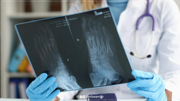
Managing Bone Fractures in Duchenne muscular dystrophy (DMD) is a critical component of multidisciplinary care, as individuals with DMD face significantly increased fracture risk due to progressive muscle weakness, reduced mobility, and glucocorticoid therapy. Fragility fractures—especially of the long bones—can lead to severe complications, including permanent loss of ambulation, chronic pain, and life-threatening conditions such as fat embolism syndrome.
Understanding fracture prevention, early recognition, and optimal treatment strategies is essential for improving long-term outcomes and quality of life in individuals living with DMD.
Table of Contents
Understanding Managing Bone Fractures in Duchenne Muscular Dystrophy
Understanding managing bone fracture in Duchenne muscular dystrophy involves a multidisciplinary, risk-adapted approach that addresses both the fracture itself and the underlying bone fragility.

Individuals with Duchenne are predisposed to fractures due to osteoporosis, long-term corticosteroid use, and reduced mechanical loading from muscle weakness. Effective management begins with prompt recognition and emergency evaluation, followed by a tailored treatment plan that may include surgical fixation for unstable fractures or casting and splinting for stable cases.
Early mobilization is prioritized—especially in ambulatory patients—to prevent permanent loss of function. At the same time, long-term strategies such as regular bone health monitoring (DEXA scans and spine imaging), optimization of calcium and vitamin D intake, and bisphosphonate therapy play a critical role in reducing future fracture risk.
Close coordination between neuromuscular specialists, orthopedic surgeons, and endocrinologists is essential to ensure safe recovery and preserve quality of life. Learn More: Bone & Joint Health in Duchenne
Why Are Bone Fractures Common in Duchenne Muscular Dystrophy?
Pathophysiology of Bone Fragility in DMD
Bone fragility in DMD results from a multifactorial process involving:
Chronic Muscle Weakness
Progressive muscle degeneration reduces mechanical loading on bones, leading to decreased bone mineral density (BMD). Mechanical stress is essential for bone remodeling; its absence accelerates bone loss.
Glucocorticoid Therapy
Long-term corticosteroid use (e.g., prednisone, deflazacort) is standard in DMD care but contributes to:
- Reduced bone formation
- Increased bone resorption
- Suppressed calcium absorption
Discover More: What Are the Pros and Cons of Steroids (Cortisone) for Duchenne
Reduced Mobility and Immobilization
Loss of ambulation significantly accelerates osteoporosis due to:
- Lack of weight-bearing activity
- Increased bone resorption
Delayed Puberty and Hormonal Factors
Endocrine dysfunction (e.g., hypogonadism) further impairs bone development.
📚 Evidence:
- Ward LM, et al. (2016). Proceedings of a Parent Project Muscular Dystrophy Bone Health Workshop
- Buckner et al. (2015) highlighted vertebral fractures as underdiagnosed complications in DMD.
Common Types of Fractures in Duchenne
Long Bone Fractures
The most frequently affected bones include:
- Femur (thigh bone)
- Tibia
- Fibula
These fractures often occur with minimal trauma due to underlying osteoporosis.
Vertebral Compression Fractures
Often asymptomatic but clinically significant:
- May present with back pain
- Can lead to spinal deformities
- Strongly associated with steroid use
📚 Study Insight: Vertebral fractures may occur in up to 30–50 percent of steroid-treated DMD patients (Ma et al., 2017).
Recognizing a Fracture in Duchenne Muscular Dystrophy
Key Signs and Symptoms
Caregivers and clinicians should monitor for:
- Severe or persistent pain
- Swelling and bruising
- Limb deformity or shortening
- Sudden loss of mobility
- Refusal to bear weight
In non-ambulatory individuals, signs may be subtle, including discomfort during transfers.
What Should I do if I Suspect a Femur Fracture?
If you suspect a femur fracture, act immediately and treat it as a medical emergency. Avoid moving the leg, keep it supported in its current position, and seek urgent care at the nearest emergency department. Do not attempt to straighten or bear weight on the limb, as this may worsen the injury. Inform the medical team about the underlying condition and contact your neuromuscular specialist as soon as possible. Early evaluation and coordinated care are essential to reduce complications such as loss of mobility, improper healing, or fat embolism syndrome.
Emergency Response to Suspected Fractures
Immediate Steps
If a fracture is suspected:
- Seek emergency medical care immediately
- Avoid unnecessary movement
- Inform the neuromuscular specialist
- Provide emergency medical documentation
Why Rapid Intervention Matters
Delayed treatment increases the risk of:
- Fat embolism syndrome (FES)
- Infection
- Permanent functional decline
Learn More: Emergency Care in Duchenne
Fat Embolism Syndrome (FES): A Critical Complication
What Is FES?
FES occurs when fat droplets enter the bloodstream following bone trauma.
Symptoms of FES
- Respiratory distress
- Confusion or altered consciousness
- Petechial rash
Clinical Importance
Although rare, FES is potentially fatal and requires urgent intervention.
📚 Reference:
- Gupta et al. (2018) emphasized early recognition of FES in neuromuscular disorders.
Learn More: Fat Embolism Syndrome in Patients with Duchenne
Treatment Options for Bone Fractures in DMD
Treatment must be individualized and coordinated between orthopedic surgeons and neuromuscular specialists.
Surgical Management
Indications
- Displaced fractures
- Unstable fractures
- Need for early mobilization
Techniques
- Intramedullary rods
- Plates and screws
Benefits
- Faster mobilization
- Reduced risk of functional decline
Risks
- Anesthesia complications
- Increased perioperative risk in DMD
📚 Evidence: Surgical fixation improves the likelihood of maintaining ambulation (Bushby et al., 2010).
Discover More: Surgery Under Anesthesia
Non-Surgical Management
Casting
- Full-leg cast (neutral alignment recommended)
- Can be weight-bearing if feasible
Splinting
- Suitable for stable fractures
- Often used in non-ambulatory patients
Rehabilitation After Fracture
Early rehabilitation is critical:
- Physical therapy
- Gradual weight-bearing
- Prevention of contractures
Multidisciplinary care is essential to optimize recovery.
Why Walkers Are NOT Recommended in DMD
Use of walkers is discouraged due to:
- Altered center of gravity
- Increased fall risk
- Upper limb weakness
Safer alternatives include:
- Wheelchairs
- Assisted mobility devices
Learn More: When Is The Best Time for a Power Wheelchair in DMD?
Preventing Bone Fractures in Duchenne Muscular Dystrophy
Fall Prevention Strategies
For Ambulatory Individuals
- Keep pathways clear
- Wear proper footwear
- Avoid overexertion
For Non-Ambulatory Individuals
- Safe transfer techniques
- Wheelchair safety belts
- Supervised positioning
Monitoring Bone Health
DEXA Scans
- Annual screening after steroid initiation
- Measures bone mineral density
Spine Imaging
- Lateral lumbar X-rays
- Genant scoring for vertebral fractures
📚 Guidelines:
Role of Endocrinology
Endocrinologists play a key role in:
- Diagnosing osteoporosis
- Managing hormonal deficiencies
- Prescribing bone-protective therapies
Medical Management of Osteoporosis in DMD
Calcium and Vitamin D
Essential for:
- Bone mineralization
- Preventing deficiency
Bisphosphonate Therapy
Mechanism
- Inhibits bone resorption
- Increases bone density
Administration
- Oral or intravenous
Benefits
- Reduces fracture risk
- Improves vertebral integrity
📚 Evidence:
- Ward LM, et al. (2016). Proceedings of a Parent Project Muscular Dystrophy Bone Health Workshop
Long-Term Impact of Fractures in DMD
Fractures can result in:
- Permanent loss of ambulation
- Increased dependency
- Reduced quality of life
- Higher morbidity
Early intervention and prevention are therefore critical.
Multidisciplinary Approach to Fracture Management
Optimal care requires coordination among:
- Neuromuscular specialists
- Orthopedic surgeons
- Endocrinologists
- Physiotherapists
This integrated model improves outcomes and reduces complications.
Read More: Multidisciplinary Neuromuscular Team in Duchenne
Future Directions in Bone Health Management
Emerging strategies include:
- Novel bone anabolic therapies
- Improved steroid-sparing treatments
- Gene therapies addressing disease progression
Ongoing research aims to reduce fracture burden in DMD populations.
FAQ: Bone Fractures in Duchenne Muscular Dystrophy
What causes bone fractures in Duchenne muscular dystrophy?
Fractures are mainly caused by osteoporosis due to steroid use and reduced mobility.
How common are fractures in Duchenne?
Up to 60 percent of patients experience fractures, especially in long bones.
What is the most serious fracture in Duchenne?
Femur fractures are particularly serious due to the risk of permanent loss of ambulation.
Should all fractures in Duchenne be treated surgically?
No. Treatment depends on fracture type, stability, and ambulation status.
Can fractures be prevented in Duchenne?
Yes. Monitoring bone health, preventing falls, and using medications like bisphosphonates help reduce risk.
What is fat embolism syndrome?
A rare but serious complication where fat enters the bloodstream after a fracture, requiring emergency care.
Are DEXA scans necessary?
Yes. Regular scans help detect bone loss early and guide treatment.
Do vitamin D and calcium help?
Yes. They are essential for maintaining bone strength and reducing fracture risk.
Final Thoughts
Managing bone fractures in Duchenne muscular dystrophy requires proactive monitoring, rapid response to injuries, and individualized treatment strategies. With appropriate multidisciplinary care, many complications can be prevented or minimized. Strengthening bone health, preventing falls, and optimizing treatment decisions are key pillars of long-term care. As research advances, improved therapies may further reduce fracture risks and enhance quality of life for individuals with DMD.
Sources and Scientific References
- Bushby K, et al. (2010). Diagnosis and management of Duchenne muscular dystrophy. The Lancet Neurology.
- Ward LM, et al. (2016). Proceedings of a Parent Project Muscular Dystrophy Bone Health Workshop
- Buckner JL, et al. (2015). Vertebral fractures in Duchenne muscular dystrophy. Neurology.
- Ma J, et al. (2017). Bone health in Duchenne muscular dystrophy. Neuromuscular Disorders.
- Birnkrant DJ, et al. (2018). DMD Care Considerations. The Lancet Neurology.
- Gupta A, et al. (2018). Fat embolism syndrome review.