Duchenne muscular dystrophy (DMD) is a severe X‑linked genetic disorder characterized by absence or dysfunction of dystrophin, a cytoskeletal protein essential for maintaining muscle fibre integrity. The disease leads to repeated muscle damage, inadequate repair, progressive fibrosis, loss of muscle mass and function, and eventually cardiopulmonary failure. Current therapies (gene therapy, exon skipping, corticosteroids, etc.) are promising but incomplete. One of the key challenges is that in humans, muscle regeneration cannot keep up with muscle degeneration, and scar formation/fibrosis worsens outcomes.
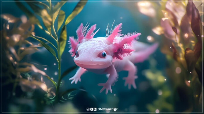
In contrast, certain animals—especially salamanders like the axolotl—display remarkable regenerative capacities. They can regrow limbs, repair internal organs, regenerate the spinal cord, and do so in a largely scar‑free way. Understanding how these processes work at cellular, molecular, and systemic levels may reveal new therapeutic angles for DMD.
Table of Contents
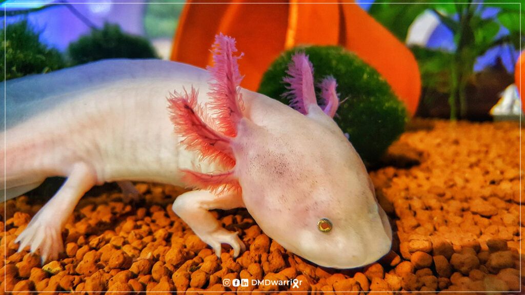
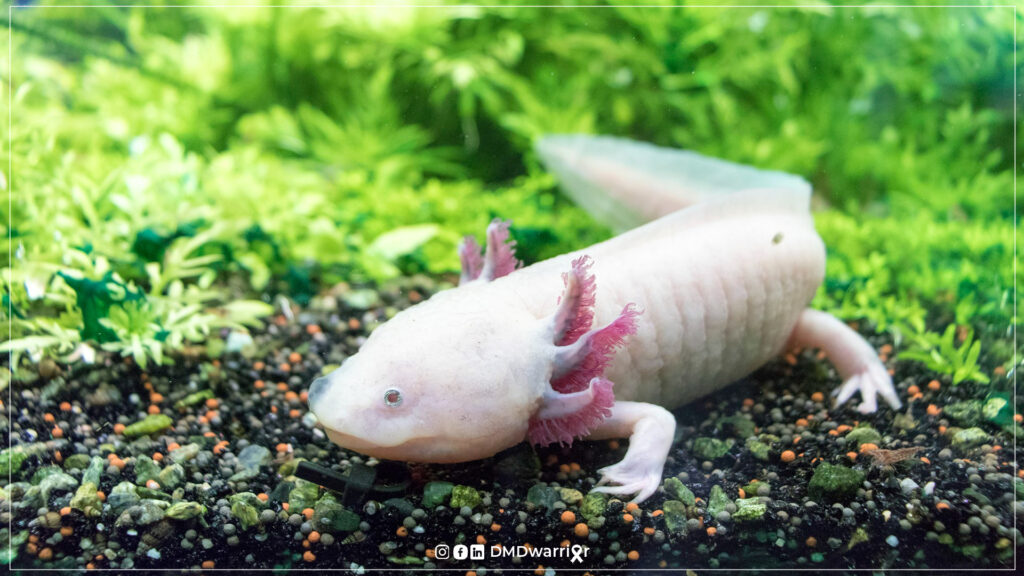
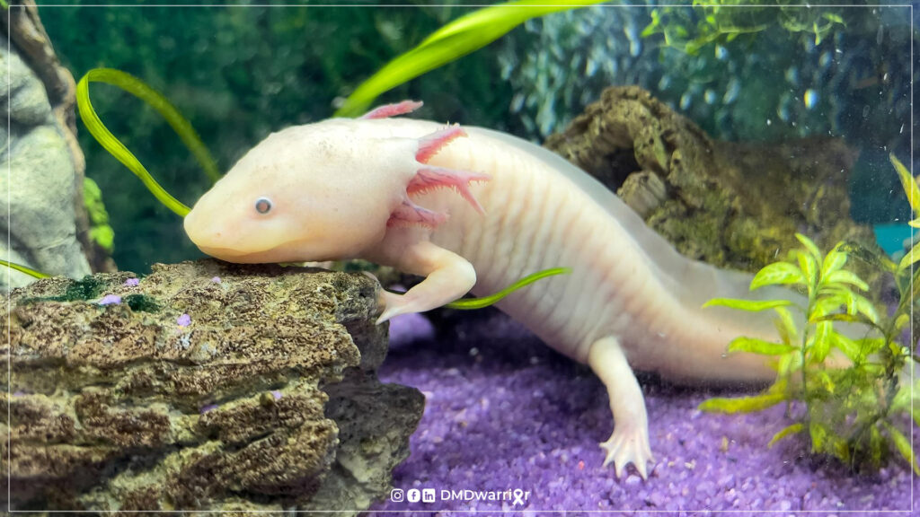
The Axolotl: Why It’s Special
Some of the key regenerative features of axolotls:
- Scar‑free regeneration of limbs, tissues, central nervous system (CNS), internal organs. –Anatomy Publications
- Ability to regenerate muscle tissue, including re‑differentiation of muscle fibres, reinnervation, rebuilding of blood vessels and extracellular matrix, and restoration of function. (There are detailed studies of limb muscle regeneration, including in hindlimbs, with proper alignment, etc.) –PubMed
- Strong, regulated growth signals during and after regeneration, including control of positional identity (e.g. via retinoic acid gradients), regulation of protein synthesis (e.g. mTOR pathway sensitivity), activation of developmental and stem/progenitor cell programs, modulation of immune response, etc. –Stanford Medicine
- Low incidence of cancer and high resistance to carcinogens. Some cell‑extracts from axolotls appear to reverse tumour growth in certain contexts. These suggest that regenerative proliferation is well controlled. –Anatomy Publications
These attributes turn the axolotl into a powerful model for understanding why regeneration succeeds in some vertebrates but fails in others (including humans), especially in contexts where there is chronic damage.
What We Know from Current Models of DMD and Where Gaps Exist
To connect axolotl insights to DMD, it helps to understand what current models show:
- The mdx mouse is the classic DMD model: it lacks functional dystrophin, shows muscle degeneration and regeneration, but the disease course is much milder than in human DMD: less fibrosis, more effective regeneration, near normal lifespan in many cases. –BioMed Central
- Larger animal models (dogs, rats) tend to show more severe phenotypes, including fibrosis, cardiomyopathy, etc., more closely resembling human disease. –PubMed
- Studies have shown not just dystrophin loss but downstream dysfunction: satellite cell exhaustion or impairment, altered gene expression in myoblasts (e.g. MyoD, Myog, etc.), increased inflammation, defective energy metabolism. –OUCI
Gaps & challenges:
- In human DMD, regenerative capacity is limited and over time fails to keep up with degeneration; fibrosis replaces muscle tissue.
- Scar formation and mis‑regulated immune responses are major obstacles.
- Many animal models don’t replicate all aspects of human disease (e.g., heart involvement, respiratory decline, long‑term outcomes).
- Translating findings from animal models to humans has been slow; gene size, immunogenicity, delivery, etc., are hurdles.
How Axolotl Research Might Help DMD
Here are ways in which the study of axolotls may contribute to new clinical research, therapies, or understanding for DMD:
| Area of insight | How axolotl research applies | Potential translational benefit for DMD |
|---|---|---|
| Muscle regeneration without fibrosis / scarless repair | Axolotls regenerate muscle tissue in limbs with low scarring; the wound epithelium forms rapidly, there is blastema formation, controlled inflammation, precise remodelling. –Wiley Online Library | Understanding the signals that suppress fibrosis and promote regenerative scarless healing may guide therapies to reduce fibrosis in DMD (e.g. targeting ECM remodelling, fibroblast activation, immune modulation). |
| Positional identity & controlled regrowth | Recent work shows that gradients of retinoic acid and enzymes that degrade it (CYP26B1) are crucial in telling regenerating cells where they are (proximal vs distal) in axolotl limbs. Genes like shox are involved in this positional identity. –NSF – National Science Foundation | For DMD, while limb patterning per se may be less directly relevant, understanding positional memory may help in aligning regenerating fibers, reconstructing architecture properly, guiding stem/progenitor cell therapies to restore correct muscle geometry. |
| Activation of developmental and stem/progenitor programs | In axolotls after injury, early upregulation of genes associated with pluripotency, cell cycle, cytoskeletal rearrangement, etc. Also the ultra‑sensitive mTOR switch (axolotl mTOR) allowing more flexible activation of protein synthesis in response to injury. –Stanford Medicine | Could suggest targets for boosting regenerative capacity in human muscle (e.g., enhancing satellite cell activity, controlling protein synthesis, metabolic programming). Perhaps treatments to “reawaken” developmental pathways that become muted in DMD due to chronic damage or aging. |
| Immune response and inflammation | Axolotls manage wound healing with an immune response that supports regeneration rather than chronic damage. Transcriptional comparisons between axolotls and non‑regenerative species after spinal cord injury highlight differences in immune pathway engagement. –MDPI | In DMD, chronic inflammation contributes to damage and fibrosis. Understanding how regeneration‑compatible inflammation is resolved in axolotls might guide anti‑inflammatory or immunomodulatory treatments that assist muscle repair rather than hinder it. |
| Molecular regulators of regeneration | Studies have identified candidate genes more highly expressed during axolotl regeneration (e.g. FSTL1, ADAMTS17, GPX7, CTHRC1) that are conserved in vertebrates. Also mechanisms like mTOR sensitivity, retinoic acid gradients. –PubMed | These conserved regulators may become drug targets: if their expression or activity can be modulated in human muscle (or in satellite cells) to enhance regeneration, one might slow disease progression in DMD. For example, molecules that mimic or upregulate FSTL1 etc. |
| Tissue scaffolding & ECM dynamics | Axolotl limb regeneration involves coordinated ECM breakdown and reconstruction (e.g. metalloproteinases, fibroblasts, etc.) that allow blastema formation and regrowth. Studies of long bones in axolotl have also used scaffolds + growth factors to promote regeneration across large defects. –National Library of Medicine | In DMD, muscle architecture is disrupted; fibrosis and ECM stiffening are problems. Insights into ECM remodelling, scaffold design from axolotl models could inform biomaterials or cell delivery scaffolds to support regeneration in dystrophic muscles. |
| Transgenesis & gene regulatory insights | Axolotl transgenic methods have improved; researchers can manipulate genes to see effects on regeneration (knock‑outs, reporter lines). –Anatomy Publications | Translating gene regulatory networks from axolotl studies could help identify human homologues or pathways for intervention; may also help validate gene therapy targets (e.g. non‑dystrophin genes that help with regeneration or ameliorate damage). |
Possible Research Strategies & Challenges in Applying Axolotl‑Derived Insights to DMD
While the potential is high, there are important considerations and steps needed for translation:
- Homology and conservation
- For any gene/pathway identified in axolotls, one must check whether there is a functional counterpart in humans (or mammals) and whether the regulatory context is similar.
- Some genes are highly conserved; others may be diverged in regulation, dosage, interaction partners. Studies like axolotl transcriptomics (blastema vs aged limbs) have started to uncover conserved gene sets. –PubMed
- Modeling chronic damage
- DMD involves ongoing cycles of degeneration/regeneration, chronic inflammation, oxidative stress, etc. The axolotl’s model of acute injury/regeneration may differ. Bridging that gap requires models with repeated injury, or sustained stress, to see whether regenerative pathways can be kept active over time.
- Scaling, strength and functional outcomes
- It’s one thing to regenerate tissue; another to restore strength, force output, neuromuscular junction stability, correct fiber type, vascular supply, etc. Translational work would need to test not just histology but functional metrics in mammalian (or possibly human) tissues or organs.
- Immunology and fibrosis
- Humans (and mammalian models) tend to develop fibrosis after injury; axolotls do not (or to a much lesser extent). Identifying what prevents fibrotic scarring in axolotls (immune suppression/regulation, fibroblast behavior, ECM remodeling) is crucial—but human immune system is more complex, with more chronic activation, etc.
- Delivery and safety
- Even if you find molecules (small molecules, proteins, gene‑therapy vectors) that activate axolotl‑like regenerative pathways, delivering them safely and effectively to human muscle (or other required tissues) in DMD patients (especially over large body areas) is nontrivial. Also, long‑term safety: regeneration often involves cell proliferation (risk of tumors), mispatterning, etc.
- Ethical and practical limitations
- The axolotl is an amphibian; though vertebrate, it is evolutionary distant. There are differences in body temperature, metabolism, scale, lifespan, etc. Translating findings will always involve dealing with species differences.
- Accessibility of axolotl lines, transgenesis techniques, and infrastructure can limit how broadly they have been used (though work is increasing). –Anatomy Publications
Specific Hypotheses & Research Proposals: How One Could Use Axolotl Insights Directly in DMD Research
Here are some concrete research ideas (hypotheses + experiments) that could exploit axolotl findings to help DMD:
- Hypothesis: One or more of the conserved genes upregulated during axolotl blastema formation (e.g. FSTL1, ADAMTS17, GPX7, CTHRC1) enhance mammalian satellite cell function under dystrophic conditions.
Experimental plan: Using cell culture, test overexpression (or recombinant protein treatment) of human homologues of these genes in dystrophic myoblasts (from mdx mice or human DMD patients) to see if proliferation, differentiation, survival, and resistance to damage are improved. - Hypothesis: Modulation of retinoic acid signalling (or CYP26B1) can influence muscle regeneration and reduce fibrotic mispatterning in dystrophic muscle.
Experimental plan: Use small‑molecule modulators of retinoic acid synthesis or degradation in mdx mice or rat models, examine not just muscle regeneration but architecture, fibrosis, NMJ integrity, functional strength. - Hypothesis: mTOR sensitivity (e.g. axolotl’s ultra‑sensitive mTOR) is part of what allows robust protein synthesis after injury; augmenting mTOR pathway in a controlled way may help dystrophic muscle.
Experimental plan: Compare activity of mTOR pathway in dystrophic vs normal muscle after injury; test mTOR activators or modulators in small or large animal models, carefully monitoring for adverse effects (e.g. maladaptive hypertrophy or metabolic stress). - Hypothesis: Immune modulation mimicking axolotl’s response may support better regeneration in DMD.
Experimental plan: Study immune cell infiltration and cytokine profiles in axolotl regeneration vs in DMD; identify molecules that correlate with resolution of inflammation (e.g. specific interleukins, growth factors). Then test blockers or enhancers of those in animal DMD models. - Use of hybrid or ‘regeneration scaffold’ materials: Based on axolotl ECM/regenerative scaffold studies (e.g. bone defect repair using scaffolds + growth factors) to design implants or scaffolds for muscle repair in DMD (e.g. filler scaffolds that degrade and allow satellite cell migration).
- Genetic/transgenic tools: Improve transgenic axolotl lines to express dystrophin homologues (if present) or model dystrophin deficiency in axolotl (if feasible) → though may be difficult, could help in understanding how certain developmental programs are suppressed or altered when dystrophin is missing.
Could Axolotls Be Used as a Model of Dystrophin Deficiency?
One question is: is there any research that uses axolotls with dystrophin deficiency (or knock‑down/knock‑out of a dystrophin‑like protein) to model DMD directly?
- From the literature We found, We did not see current, published work showing that axolotls have been genetically modified to lack a dystrophin orthologue, or used as a direct DMD model. The standard DMD models remain mammals (mdx mice, DMD rats, canine models).
- However, studies have identified many homologues in axolotls of human proteins implicated in regenerative biology and disease (e.g. in neurodegeneration). This suggests that many of the “toolkit” genes are present. –MDPI
- The transgenic toolkit in axolotls is improving, so if scientists can introduce mutations or knockdowns for dystrophin (or a dystrophin‑like gene), then one could observe how regenerative mechanisms behave in absence of dystrophin, and whether axolotl’s regeneration can compensate or bypass some of the deficits.
Thus, while direct axolotl DMD models are not yet established (as far as the literature We surveyed), there is potential for such models to be developed.
What to Watch for / Key Metrics if You Were Designing Axolotl‑Inspired DMD Therapies
To ensure translational relevance, research would need to evaluate:
- Muscle histology: fibre size, central nuclei, necrosis/regeneration cycles, fibrosis, fat infiltration.
- Functional strength: force generation, endurance, contractile properties.
- Neuromuscular junction (NMJ) integrity: since DMD also affects NMJs via cycles of degeneration/regeneration.
- Inflammation / immune cell infiltration and resolution over time: both acute and chronic markers.
- Extracellular matrix (ECM) changes: collagen deposition, stiffness, scaffold properties.
- Molecular signatures: expression of developmental/regenerative genes, protein synthesis capacity (mTOR etc.), metabolic status (mitochondria, reactive oxygen stress).
- Side effects: risk of mispatterning, tumourigenesis, off‑target effects, immune reactions.
Challenges & Limitations
- Species differences: The environment of regeneration (temperature, metabolism, lifespan, immune system) is different in amphibian vs mammalian context. Some mechanisms may not translate or may work differently.
- Chronic vs acute injury: Axolotl studies often examine acute injury/regeneration, not the chronic, accumulating damage seen in DMD. If regenerative programs get “used up” or are inhibited in chronic settings, findings from axolotls may lose potency.
- Delivery in humans: If the target is to enhance regeneration via small molecules, growth factors, gene therapy, cell therapy, etc., then delivering these safely and effectively to the large muscle masses in humans is hard. DMD is systemic; it involves many muscles.
- Timing: Interventions in DMD likely need to be early (before massive fibrosis and loss of satellite cells). Many axolotl events happen immediately after injury; in DMD patients, often damage and fibrosis are present long before diagnosis and therapy.
- Regulation and safety: Any therapy that promotes cell proliferation (to mimic regeneration) also raises risk of cancer or unwanted tissue growth if not tightly controlled.
Conclusion
The axolotl has a remarkable regenerative toolkit, involving scarless repair, precise control of growth and positional identity, efficient activation of developmental pathways, robust modulation of immune response, and highly regulated ECM remodeling. While direct models of DMD in axolotls are not yet established, the conserved nature of many of the genes and pathways involved means that insights from axolotl research have real promise for contributing to therapies or mechanistic understanding in DMD.
Potential takeaways include:
- Identification of new molecular targets (genes, signalling pathways) to enhance regeneration or slow fibrosis.
- Development of small‑molecule or biologics that mimic axolotl regenerative cues (e.g. retinoic acid regulation, mTOR modulation).
- Designing scaffolds or biomaterials informed by axolotl ECM dynamics.
- Better understanding of how immune response can be managed to support regeneration rather than hindering it.
For clinical research, collaboration between regenerative biology (axolotl labs), DMD clinicians, mammalian model systems, and therapeutic developers will be essential. Ultimately, while axolotl‑derived strategies will likely not cure DMD alone, they may significantly improve quality of life, slow progression, enhance muscle repair, or work in combination with gene therapies or exon skipping.
Read More: Clinical Trials for Duchenne (List of All Researches)
Watch It: Salamander Limb Regeneration